Morphology, Habitat, Host, and Life Cycle of Balantidium coli
26/12/2022 Views : 38656
I K. Putra Juliantara
Morphology, Habitat, Host, and Life Cycle of Balantidium coli

Figure 1. Forms of cysts and trophozoites of Balantidium coli (CDC, 2016)
Balantidium coli has two stages, namely the trophozoite stage and the cyst stage. The trophozoite stage is 60-70 microns long and 40-50 microns wide, has a depression in the anterior part of its body called the peristome where there is a mouth (cytosome). These ciliates do not have a gut, but have an anus (cytopyge) which is in the posterior part of the body. Pictures of Balantidium coli cysts and trophozoites can be seen in Figures 1, 2 and 3 (Soedarto, 2016)

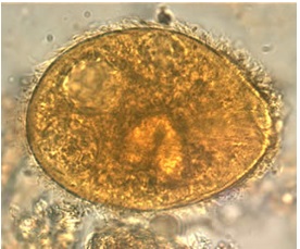
Figure 2 Cyst form of Balantidium coli (CDC, 2017)
In the anterior part which is rather narrow, there is a cytostome which functions as a mouth. The posterior part of the shape is slightly widened, in this area a cytopage (cytophyge) is found which functions to remove substances that are no longer needed. On the entire surface of the body there are vibrating hairs (cilia) arranged in longitudinal rows. On the cytostome there are rather long vibrating hairs. The function of the vibrating feathers is to move and take food (Lecturer Staff of the Department of Parasitology, 2008).

Figure 3 Trophozoite form of Balantidium coli (CDC, 2017)
Balantidium coli has two nuclei, namely a large, kidney-shaped macronucleus and a micronucleus which is shaped like a small dot which is in the hollow part of the macronucleus. Trophozoites have two contractile vacuoles and several food vacuoles which contain food debris, leukocytes and erythrocytes. The structure of Balantidium coli can be seen in Figure 4 (Soedarto, 2016).

Figure 4 Trophozoite Structure and Cysts of Balantidium coli (Jain, 2016)
The shape of the parasitic cyst is round, measuring in diameter between 50 and 60 microns, has a two-layer cyst wall. The cyst has a granular cytoplasm containing a macronucleus, a micronucleus and a retractile body. Contractile vacuoles can still be found occasionally. This parasite lives in the mucous membrane of the large intestine, especially in the cecum area (Lecturer Staff of the Department of Parasitology, 2008; Soedarto, 2016).
The habitat of Balantidium coli in humans is the cecum and colon. Humans are infected with Balantidium coli by ingesting water or raw food contaminated with pig feces containing infective cysts of this parasite. So, the natural habitat of Balantidium coli is in pigs (Schuster and Avila, 2008; Soedarto, 2016). Balantidium coli is a cosmopolitan opportunistic parasitic pathogen that can be found worldwide. This ciliate infection was reported from various countries, especially those where many people raise pigs. These organisms are cosmopolitan and can be found anywhere pigs are found. Balantidiosis appears to be a problem in most developing countries, where water sources may be contaminated with pig or human feces. Balantidium coli can become an opportunistic parasite in immunosuppressed hosts living in urban environments, where pigs are not a factor in infection (Schuster and Avila, 2008; Soedarto, 2016).
Hosts of this parasite include humans, pigs, dogs, primates and rats that live in the tropics. Pigs are the main source of transmission of balantidiosis for humans because these animals are the definitive natural hosts and also act as reservoir hosts for humans which are actually only incidental hosts for this parasite (Lecturer Staff of the Department of Parasitology, 2008; Soedarto, 2016).
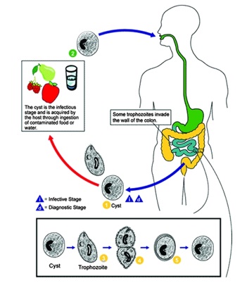
In the life cycle of Balantidium coli, the cyst stage and the trophozoite stage can occur in one type of host (figure 5). Pigs are the main source of transmission of balantidiosis for humans because these animals are the definitive natural hosts and also act as reservoir hosts for humans which are actually only incidental hosts for this parasite (Soedarto, 2016).
Humans are infected with Balantidium coli by ingesting water or raw food contaminated with pig feces containing infective cysts of this parasite. In the large intestine, the cyst turns into a trophozoite form which then grows and reproduces by means of cell division (binary transverse fission) or by conjugation in the intestinal lumen or in the intestinal submucosa. Picture of the life cycle of Balantidium coli can be seen in Figure 2.5 (Soedarto, 2016).
The vegetative stage is also the stage that functions to reproduce by means of transverse pairs. First, the micronucleus divides, followed by the macronucleus and cytoplasm to form two new organisms. Sometimes there is an exchange of chromatin (conjugation). Trophozoites will immediately form cysts (encystation) in the intestinal lumen or immediately after being excreted in the stool. Conjugation reproduction occurs by means of two trophozoites forming a cyst together, then exchanging core material, finally splitting back into two new trophozoites. If the environment in the intestine is not suitable for parasite life, the trophozoite will turn into a cyst form (Lecturer Staff of the Department of Parasitology, 2008; Soedarto, 2016).

Figure 5 Life cycle of Balantidium coli (Schuster and Avila, 2008)
The cyst, approximately 60 microns in size, is oval and thick-walled. Cysts have only a macronucleus. A living cyst has vibrating hairs that are still moving. Cysts are not for breeding, their function is only to survive. Cysts in the stool can survive 1-2 days at room temperature. Cysts are the infective form. If the cyst is swallowed, excystation occurs in the small intestine. From one cyst comes out a vegetative stage which immediately multiplies and forms colonies in the mucous membrane of the large intestine. The cyst stage and the vegetative stage are excreted with the host's feces. Infection occurs when cysts are swallowed (Teaching Staff of the Department of Parasitology, 2008).
REFERENCES
Centers for Disease Control and Prevention. 2016. Parasites – Balantidiasis. [serial online]. [cited on 20 Des 2022]. Available from: https://www.cdc.gov/parasites/balantidium/.
Jain, K. 2016. Example of Phylum Protozoa: Balantidium coli. [serial online]. [cited on 20 Des 2022]. Available from: http://www.biologydiscussion.com/animals-2/phylum-protozoa/example-of-phylum-protozoa-balantidium-coli/32461
Schuster, F.L. and L. R. Avila. 2008. Current World Status of Balantidium coli. [serial online]. [cited on 20 Des 2022]. Available from: https://www.ncbi.nlm.nih.gov/pmc/articles/PMC2570149/
Soedarto. 2016. Buku Ajar Parasitologi Kedokteran. Jakarta: Sagung Seto
Staf Pengajar Departemen Parasitologi. 2008. Buku Ajar Parasitologi Kedokteran. Jakarta: Badan Penerbit FKUI
