Morphology, Geographic Distribution, and Life Cycle of Acanthamoeba sp.
29/07/2023 Views : 477
I K. Putra Juliantara
Like other
amoeba, Acanthamoeba sp. Sp. It
consists of ectoplasm and endoplasm. Inside the endoplasm is a vesicular
nucleus with a large karyosome and a nuclear wall full of chromatin grains; In
addition to the core, there are also food vacuoles. In the genus Acanthamoeba sp. Tropozoites have varied
shapes. The cyst stage is about 20 μm in size, has a round or oblong shape. The
cyst has a double wall, the outer side of which is wrinkled (Sutanto et al,
2013).
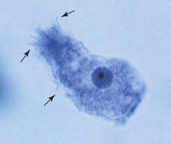
In Acanthamoeba sp., the tropozoites have size around 25 μm, (Figure 1). The motility of these tropozoites is slow, with limited evidence of progressive movement. Each tropozoite contains a single nucleus, characterized by large karyosomes similar to those found in Naegleria fowleri. However, clear peripheral chromatin is not present. The cytoplasm of the tropozoites appears granular and undecomposed (Zeibig, 2013).

Figure 1 Tropozoite
Acanthamoeba sp.
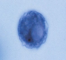
The cysts of the species Acanthamoeba sp. are spherical in shape and measure between 8μm to 25μm (Figure 2). Each cyst is encased in a double cell wall, with a smooth inner cell wall surrounded by a jagged outer cell wall. This distinctive feature is characteristic and facilitates the identification of Acanthamoeba sp. cysts. The single nucleus within the cyst resembles the shape of the corresponding trophozoite, with a large karyosome and the absence of prominent peripheral chromatin (Zeibig, 2013).

Figure 2 Acanthamoeba sp. cyst
Acanthamoeba sp. is one of the most commonly found species
in nature, distributed worldwide, and has been isolated from various sources
including soil, dust, air, natural and treated fresh water (such as swimming
pools and warm baths), seawater, air conditioners, fecal waste, sediment,
hospitals, contact lenses, and agricultural crops. It has also been found in
plants, various animal species including fish, amphibians, reptiles, and
mammals, as well as in the nasopharyngeal cavity of apparently healthy
individuals and those with immunodeficiencies. Acanthamoeba sp. can thrive
within a broad temperature range in water, with its optimal growth temperature
at 30°C. The trophozoite stage of Acanthamoeba sp. is capable of living and
replicating in water, feeding on bacteria, yeast, and other microorganisms.
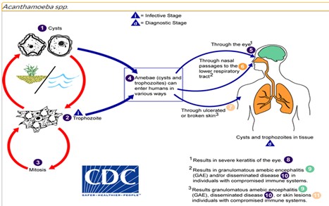
In the life cycle of Acanthamoeba sp., there is no flagellum stage present. The life cycle can be observed in Figure 3. Trophozoites replicate through mitosis, with the nuclear membrane not remaining intact. Although trophozoites are an infective form, cysts and trophozoites can enter the body through various routes. Entry can occur through the eyes, nasal passages, lower respiratory tract, or through ulcerated or broken skin. When Acanthamoeba sp. infects the eye, it can cause severe keratitis in healthy individuals, particularly among contact lens users. If it enters the respiratory system or the skin, it can invade the central nervous system through hematogenous spread, leading to granulomatous amebic encephalitis (GAE) (Qvarnstrom, 2017).

Figure 3
Life Cycle of Acanthamoeba sp.
REFERENCES
Qvarnstrom. Y, G. S. Visvesvara., R. Sriram., A.J. da
Silva. 2017. Multiplex real-time PCR assay for simultaneous detection of Acanthamoeba sp. spp, Balamuthia mandrillaris, and
Naegleria fowleri.
J Clin Microbiol. CDC Journal, 44
(10): 3589-3595.
Sutanto. I., I. S. Ismid., P. K. Sjarifuddin., S.
Sungkar. 2013. Textbook of Medical
Parasitology fourth edition. Jakarta: FKUI Issuing Agency.
Zeibig.
E. 2013. Clinical Parasitology: A Practical Approach
Second Edition. St. Louis,
Missouri:
Elsevie.
