Radiological Imaging in Osteomyelitis: Early Detection and Diagnostic Challenges
14/12/2024 Views : 90
Faradilla Novita Anggreini

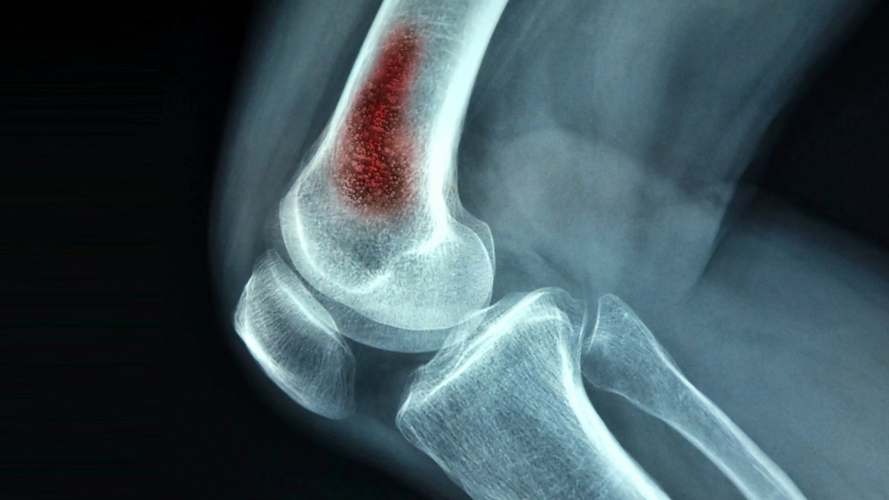
Osteomyelitis is a bone infection caused by bacteria or fungi, often affecting children, individuals with weakened immune systems, and patients with certain medical conditions such as diabetes. Early diagnosis is crucial to prevent serious complications, such as bone necrosis and sepsis. In the field of radiology, various imaging modalities are used to detect and evaluate osteomyelitis, ranging from conventional radiography to advanced imaging techniques like MRI and CT scans.
Conventional radiography is commonly used as an initial examination for detecting osteomyelitis. However, signs of acute osteomyelitis, such as soft tissue swelling, may not be visible within the first 7 to 10 days of infection. In later stages, bone changes like cortical lysis and periosteal reactions begin to appear. Computed Tomography (CT) scans provide more detailed images of bone structure and are particularly useful for detecting bone destruction and subperiosteal abscesses that may form due to the infection. Magnetic Resonance Imaging (MRI) is the best modality for detecting osteomyelitis in its early stages, as it clearly shows bone marrow edema and abscess collections. Additionally, MRI is useful in distinguishing infections from non-infectious processes such as tumors or trauma. Nuclear imaging techniques, such as bone scans and PET scans, also play an important role in diagnosing osteomyelitis. Bone scans use radioisotopes like technetium-99m to detect increased metabolic activity in infected bones, while PET scans with fluorodeoxyglucose (FDG) can identify areas of inflammation with high sensitivity.
Although radiological imaging plays a critical role in diagnosing osteomyelitis, there are several challenges that need to be addressed. One of these challenges is that radiological changes often appear later than the clinical progression of the disease itself. Additionally, osteomyelitis can resemble other conditions, such as bone tumors, septic arthritis, or trauma, requiring further confirmation with advanced imaging techniques. In patients with orthopedic implants, artifacts from the implants may interfere with imaging, especially in MRI scans.
Overall, radiological imaging plays a vital role in detecting osteomyelitis, particularly in identifying infections at an early stage and helping determine appropriate treatment strategies. The combination of various imaging modalities, such as radiography, MRI, CT scans, and bone scans, can enhance diagnostic accuracy and minimize the risk of more severe complications.
